
In this Article
Palpitations
How Are Palpitations Diagnosed?
When you tell your doctor about the palpitations, you will likely be asked a number of questions, such as:
- How often the palpitations occur
- How long they last
- What circumstances bring on the palpitations
- Whether other symptoms occur with the palpitations.
The doctor will then perform a physical examination to look for signs of conditions that may be causing your palpitations.
Your doctor can order a variety of tests to determine whether your palpitations are due to an abnormal heart rhythm. Some of these tests evaluate the heart’s chambers, valves, and arteries. Others focus primarily on the heart’s electrical system.
|
How-To Information: Ways in which your doctor can evaluate your palpitations:
|
Routine Electrocardiography
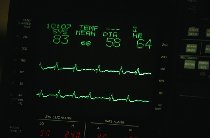
Routine or standard electrocardiography is the most commonly performed test in cardiology. This painless and harmless test produces an electrocardiogram (ECG or EKG) , which is a record of the electrical activity of the heart.
In this test, surface electrodes attached to wires (leads) are applied to the skin of your chest and limbs. These leads send your heart’s electrical signals to the electrocardiograph machine, which records this information on paper. The ECG shows a series of waves that represent the electrical events of various chambers and the conduction pathways within the heart.
A standard 12-lead ECG can diagnose disturbances in the heart rate or rhythm associated with palpitations, but only if palpitations occur while the ECG is being obtained. In addition to recording the heart’s rhythm, the ECG may help to identify an underlying cardiac condition, such as thickening of the heart muscle due to high blood pressure, disease of the heart valve, or a previous heart attack (myocardial infarction).
For further information about high blood pressure, go to high blood pressure.
For further information about heart attack, go to heart attack.
Long-Term ECG Recording
Individuals with suspected heart rhythm irregularities that are not recorded on a routine ECG will often undergo some type of home or ambulatory ECG monitoring. These methods include 24-hour Holter monitoring, transtelephonic event recording, and continuous loop recording.
- Holter monitor – A Holter monitor is a portable ECG recorder that you wear for 24 hours to obtain a continuous recording of your heart’s electrical activity. The ECG is recorded on a tape or computer chip and later analyzed by a computer for interpretation by your doctor. Several electrodes are placed on your chest and are connected by wires to a small recorder.
This recorder can be worn on a strap over your shoulder or around your waist while you continue with your normal daily activities. You will be asked to keep a diary while wearing the recorder so correlations may be made between your palpitations and the associated rhythm.
- Event (transtelephonic) recorders – If you do not feel
palpitations very often, you may be given a machine called an event recorder to keep with you. Event recorders are small devices that record short ECG intervals and are not intended to be worn all of the time.When you start to feel palpitations, you take it out and attach the device to your body temporarily. You do this by putting on a bracelet that attaches to the recorder or by pressing the device against your chest. You then press a button that initiates recording of your heart rhythm for up to one minute. The ECG is then transmitted by telephone to your doctor’s office for interpretation.
- Loop recorders – A continuous loop recorder is an ECG recorder applied to the surface of the skin that records several minutes of electrical activity at a time. It continuously records new information while discarding old information, so its memory stores only several minutes worth of information. But you can “freeze” the recording in the device’s memory if you experience palpitations.
As with event recorders, you transmit information recorded on a computer chip to your doctor’s office by telephone. These devices are useful for capturing brief episodes of electrical activity when it takes too long to apply an event recorder.
|
Nice To Know: An implantable form of a continuous loop recorder, known as an implantable loop recorder (IRL), may be implanted under the skin. These devices require no external electrodes or power source and use a small hand-held activator to “freeze” ECG information surrounding an event. |
Signal-Averaged ECG
This noninvasive test produces a surface ECG that allows your doctor to detect areas of slowed ventricular activation. This test is useful in determining whether an individual is at risk for ventricular
Exercise ECG (Stress Test)
In this test, an electrocardiograph machine records your heart’s electrical activity while you increase your heart rate by walking on a treadmill. This allows doctors to study changes in your ECG while your heart is made to work harder.
A stress test can show if the arteries that supply blood to the heart are partially blocked, as may occur with
Echocardiography
An ultrasound probe is placed on your chest and sound waves are directed through the probe to your heart. A computer shows images of the heart on a video screen by “reading” echoes of the sound waves as they bounce off the heart. This procedure does not involve any exposure to radiation and is painless.
Chest X-ray
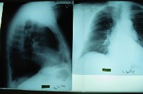
The chest x-ray is an examination that employs a beam of energy (ionizing radiation) to obtain images of the heart, lungs, and ribs. It allows a physician to view the size of the heart and may reveal underlying heart or lung problems that could be linked to the palpitations.
Blood Tests
On occasion, a sample of blood is taken for specific laboratory tests. These tests can identify or rule out chemical causes of rhythm disturbances, such as an overactive or underactive thyroid gland, anemia, kidney problems, or abnormal levels of sodium, potassium, calcium, or magnesium.
Other Tests
Some more invasive tests involve the use of thin tubes called catheters, which are inserted into a blood vessel, usually near the groin. These catheters then are guided up through the blood vessels to the heart. The following two tests are performed at the hospital and may require an overnight stay.
Cardiac catheterization provides information about the anatomic structures of the heart as well as pressures inside the heart chambers. This test is useful for diagnosing various cardiac conditions that may be associated with arrhythmias.A dye can be injected through the catheters that allow the arteries of the heart to be seen on an X-ray film and checked for blockages seen with coronary artery disease. Dye can also be injected into the heart’s pumping chambers to see how well the heart muscle is contracting and the valves are working.
- An electrophysiologic (EP) study is used when spontaneous arrhythmias are infrequent but when a serious
arrhythmia is suspected. This test involves inserting wires (electrode catheters) into leg, arm, or neckveins and guiding them into the heart using x-ray.In addition to obtaining information about electrical events from various locations in the heart, these catheters can serve as pacemakers and allow the doctor to stimulate the heart to produce a change in heart rhythm. This can allow the doctor to observe the portion of the heart involved in producing the arrhythmia. This test is useful in identifying abnormal circuits (electrical pathways) in the heart, which can then be destroyed with radiofrequency energy so the arrhythmia will not recur. The area that is destroyed is very small.