
In this Article
Macular Degeneration
What Is Age-Related Macular Degeneration (AMD)?
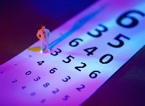
Age-related macular degeneration (AMD) is the leading cause of vision loss in people older than 55 years of age. Macular degeneration causes loss of the sharp, central vision needed for many daily tasks that involve looking straight at objects. People with AMD may find it difficult or impossible to drive, read, sew, or recognize faces. Many also have trouble distinguishing colors.

AMD results from deterioation of the central part of the retina, called the macula (MAK-yoo-luh). The retina is a tissue-thin membrane lining the back of the eye. We see objects because light passes through the eye and strikes the retina. Light-sensitive cells in the retina capture images from the outside world and relay them to the brain. The macula, which is about the size of a pencil eraser, relays images in the direct line of focus. People lose this central vision when macular cells degenerate, or stop working normally. The degeneration and vision loss usually occurs slowly, over a period of years. People with AMD retain peripheral vision, or side vision.
|
Facts about AMD
|
What Causes Macular Degeneration (AMD)?
Scientists don’t know. Factors associated with the aging process obviously play a big role, since AMD occurs as people grow older. Heredity is another factor. People with a close relative who had AMD have a greater chance of developing AMD themselves. The “wet” form (see below) is associated with smoking and high blood pressure.

Experts disagree about the role of nutrients in AMD. Some think that lack of certain nutrients in the diet, especially zinc, may increase the chances of getting AMD.
Different Forms Of Macular Degeneration
Yes. Some rare kinds of macular degeneration occur in children. Called juvenile macular degeneration, they are hereditary diseases caused by abnormal genes passed from parents to their children. Macular damage can result from injuries to the eye, exposure to intense light, and infections. People with severe myopia, or nearsightedness, may develop macular degeneration. Macular damage can occur as a complication of diabetes and certain other diseases.
Most cases of macular degeneration, however, are age-related. AMD occurs in two forms, a “dry” form and a “wet” form. Both are painless. Wet AMD also is called neovascular macular degeneration because it involves growth of new blood vessels. “Neo-” means “new” and “vascular” means blood vessels. Dry AMD is called the non-neovascular form of AMD because it does not involve growth of new blood vessels.
- Dry AMD. About 90 per cent of people with AMD have the dry form. It involves a slow breakdown of light-sensitive cells in the
retina and a gradual loss of central vision. Dry AMD may progress into the more serious “wet” form. - Wet AMD. Although only 10 per cent of the people with AMD have the wet form, it causes 90 per cent of the blindness from AMD. In wet AMD, abnormal new blood vessels grow under the
macula . The vessels have unusually delicate walls, and may begin to ooze blood and fluid. The fluid damages nearby light-sensitive retina cells. If the leaks continue, all of the cells in the macula may be injured within a few weeks.
What Are The Symptoms Of Macular Degeneration (AMD)?
In the early stages, AMD causes no symptoms. That’s one of the reasons why the American Academy of Ophthalmology recommends an annual eye examination starting at age 60 to 65 to check for AMD. The exam also can find other common conditions, including cataract and glaucoma.

Symptoms may appear in one eye, or both, and include any of the following:
- Fuzzy or blurry vision
- An empty or dark area in the center of your vision
- Straight lines, such as sides of buildings, telephone poles, or sentences on a page, appear curved or wavy
- A dimming of vision when reading
Only an eye doctor can determine if the symptoms are due to AMD, or some other condition. People who experience symptoms should make an appointment with an eye care specialist immediately. Early diagnosis is important to preserve vision.
How Is Macular Degeneration Diagnosed?
The doctor will take your medical history, ask about changes in your vision, and check your eyesight. Then you will get tests that can detect not just AMD, but other eye problems such as glaucoma and retina detachments.

The doctor will give you eye drops that enlarge, or dilate, the pupil of the eye. Dilating the pupils gives the doctor a larger opening to view the inside of the eye. He or she then will use instruments to look into your eyes and examine of the retina for signs of AMD.
One of the first signs of AMD are tiny deposits, called
The doctor also may ask you to look at an Amsler Grid, a checkerboard-like pattern of lines. If the lines appear curved or distorted, it may be a sign of early AMD.
If the doctor suspects wet AMD, he may do a test called
What Is The Treatment For Dry AMD?
There is no proven treatment for dry AMD. Doctors usually watch or monitor dry AMD for the first signs that it is progressing to the more dangerous wet AMD. People with dry AMD usually return to the eye doctor on a regular basis for checkups every 6 to 12 months.
They also may be given an Amsler Grid to take home and use between visits to the eye care professional. The individual covers one eye, and looks at the grid with the other while holding the grid at normal reading distance.
- People with normal eyesight can see the center dot, four corners, and sides of the Grid. The lines appear straight and clear.
- People with AMD see wavy, broken, or distorted lines. There may be blurred spots or holes.
By looking at the Grid every day, a person with AMD can watch for vision changes. They may be early warning signs that dry AMD is changing to wet AMD. Early treatment of wet AMD may prevent further loss of vision. If changes appear on the Grid, contact your eye care professional immediately.
Can Anything Else Be Done For Dry AMD?

Some research studies have hinted that taking certain vitamin and mineral supplements may be beneficial.
- Dietary supplements may help control dry AMD, and reduce the risk that it will change or progress to wet AMD.
- One of the dietary supplements is the mineral zinc. The
retina needs relatively large amounts of zinc to stay healthy. Many older people eat diets low in zinc, found mainly in meat, poultry and fish. - Others include
antioxidants like beta carotene, vitamin C, and vitamin E. Antioxidants may protect body tissue from age-related damage. - If you have dry AMD, ask your eye doctor about taking vitamin and mineral supplements. Many common foods are good sources of these vitamins and minerals. These include spinach, carrots, collards, and other deeply colored vegetables; citrus fruits; and whole grains.
- The
National Eye Institute is sponsoring aclinical trial to see if nutritional supplements really do stop or slow the progression of dry AMD. About 4,000 people are in the study. One-third are taking vitamins, one third vitamins and minerals, and one third are taking a pill with no medicine for comparison purposes. The study will show whether supplements are safe and work and which work best.
What Is The Treatment For Wet AMD?
If detected at an early stage, wet AMD can be treated with two methods, laser surgery or photodynamic therapy.
Laser Surgery
Laser surgery is done in a doctor’s office or clinic and takes only a few minutes. Individuals undergoing treatment may get a local anesthetic to ease any discomfort from a contact placed on the eye during treatment. The actual treatment does not hurt. They can go home right after the procedure and resume normal activities.
Laser surgery does not involve cutting into the eye. Rather, the doctor shines a laser beam through the pupil of the eye and onto the retina. The beam destroys new blood vessels that are growing in the macula.
Laser surgery does not cure AMD, or restore lost vision. It does prevent additional loss of vision. By destroying fragile new blood vessels, it prevents oozing of blood and fluid that damages macula cells. Laser surgery may be repeated if more new blood vessels start growing in the macula.
- Laser surgery is suitable for only about 15 percent of people with wet AMD.
- Most people seek treatment too late – after new blood vessels already have grown too far into the focusing area of macula.
- If done at this advanced stage, laser surgery could further damage vision.
Photodynamic Therapy (PDT)
Photodynamic therapy (PDT), was approved by the U. S. Food and Drug Administration in April 2000. It involves injecting the patient with a special drug that becomes active when exposed to a certain kind of laser light. The drug flows into the abnormal vessels in the macula. Then the eye doctor focuses the special laser on the vessels. Laser light activates the drug, causing a chemical reaction that destroys the abnormal blood vessels.
- PDT is done in the doctor’s office, and the patient can resume normal activities after treatment.
- Treatment must be repeated about 5 times over a period of two years.
- Clinical trials showed that macular degeneration remained stable and did not worsen in about 67 per cent of patients treated with PDT. In contrast, the disease remained stable in 30 per cent of control patients who got no PDT.
Are There Any Clinical Trials On New Treatments?
Clinical trials do provide people with the earliest possible access to potential new treatments for a disease. In addition to the benefits, there are potential risks of being in a clinical trial. The new treatment, for instance, may not be effective and may have unrecognized side effects.
Among the trials underway are surgery to remove abnormal vessels and anti-angiogenesis drugs to stop leaking blood vessels.
A list of clinical trials at almost 50,000 locations nationwide is available at http://www.clinicaltrials.gov. Check it regularly for new AMD clinical trials.
What Is The Outlook For Better Treatment And Prevention of AMD?
Research studies are underway on the causes, prevention, and treatment of AMD. The Federal Government has given AMD higher priority in recent years. That’s because AMD will become a more serious national health problem as “baby boomers” grow older.
- Scientists are trying to identify genes that cause AMD, or increase risk for the disease. Once AMD genes are found, scientists may use gene therapy to replace the defective genes with normal copies.
- Researchers also are working on medicines that stop the growth of new blood vessels. Such drugs could be used to treat wet AMD, and perhaps stop dry AMD from getting worse.
- The clinical trials on dietary supplements, of course, could provide a simple and inexpensive way to reduce the risk of AMD.
Macular Degeneration: Frequently Asked Questions
Here are some frequently asked questions related to macular degeneration.
Q: I’ve just been diagnosed with AMD. So far my only problem is a very slight distortion of sight in my right eye. Telephone poles and other straight-line objects look a little curved. My left eye is fine. But I’m concerned about the future. Will the disease get worse and worse until I’m completely blind?
A: AMD almost never causes total blindness. It affects mainly central vision needed for viewing objects straight ahead. People with serious loss of central vision still retain side, or peripheral, vision. They can see by turning to the side. Magnifying devices and other low-vision aids are available today. These magnifying lenses and electronic devices can improve vision in people with AMD. Remember, that’s the worst-case situation. Only about 10 percent of people with AMD have the most serious form. This “wet” AMD, which involves bleeding into the
Q: My ophthalmologist gave me a piece of paper with a Grid-like pattern of lines to take home and look at regularly. She wants me to phone immediately if any of the lines start appearing wavy or blurred. What’s the point?
A: It’s a very important point. The chart is called an Amsler Grid. You should use it to monitor your vision between visits to the doctor. People with normal vision see the center dot in the grid, and all the lines. The lines appear straight and unbroken. Call your doctor right away if holes or blurry spots appear in the grid, or if the lines appear curved, broken, or distorted. It may mean that your AMD is progressing. Quick treatment may bring the disease back under control, and minimize permanent loss of vision.
Q: Friends have told me about a new treatment for AMD called
A: Doctors have treated AMD with laser surgery for years. Despite its name, laser surgery does not involve cutting into the eyeball. During the procedure, the doctor focuses a laser beam through the
Q: My mother and grandmother both have AMD. Although I’m only 25, I’m worried about getting the disease when I’m 60 or 65. Can young people take any precautions to prevent AMD?
A: There is no scientifically proven way to prevent AMD. The risk of AMD is greater in individuals who have a close, older relative with the disease. People with a family history of AMD may want to take extra care about eating a diet with ample amounts of zinc, vitamins C and E, and beta carotene. These nutrients may help reduce the risk of AMD. Chat with your doctor about taking dietary supplements, as well. Protecting your eyes from strong sunlight also may help, especially if you have light-colored eyes. AMD is more common in fair-eyed people. Eye color may explain why AMD is relatively rare in the African-American population. When in the sun, wear eyeglasses that filter out ultraviolet (UV) rays. Avoiding cigarette smoking and environmental cigarette smoke also may help. Studies show that smokers are more apt to develop AMD.
Q: I’ve heard that drinking one or two glasses of wine each day may reduce the risks of AMD. Does wine consumption have any effect?
A: A 1998 study of 3,072 adults did find hints that people who consume small amounts of wine may be less likely to develop AMD. It was published in the May issue of the Journal of the American Geriatrics Society, and got a lot of publicity. Experts at the
Macular Degeneration: Putting It All Together
Here is a summary of the important facts and information related to macular degeneration.
- Macular degeneration is an eye disease that causes the loss of the sharp, central vision needed for many daily tasks.
- Macular degeneration most often occurs as people age, although a form of it can affect children.
- Symptoms of macular degeneration include fuzzy or blurry vision, an empty or dark area in the center of your vision, straight lines such as telephone poles or sentences on a page that appear to be wavy, and a dimming of vision when reading.
- The two types of age-related macular degeneration are dry AMD and wet AMD. Wet AMD causes most of the blindness that can result from macular degeneration.
- There is no proven treatment for dry AMD, but because it can progress to the more serious wet AMD, regular eye exams are important.
- In its early stages, wet AMD can be treated with
laser surgery or photodynamic therapy. - Laser therapy involves using a laser beam to destroy new blood vessels growing in the eye. The goal is to prevent additional loss of vision.
- Photodynamic therapy involves injecting the patient with a special drug that flows into the abnormal blood vessels in the eye. A special laser focused on these vessels causes a chemical reaction that destroys abnormal vessels.
- New surgical techniques and new medications are under investigation to treat macular degeneration.
Macular Degeneration: Glossary
Here are definitions of medical terms related to macular degeneration.
Antioxidants: Nutrients like vitamin C and vitamin E that protect cells in the body from damage that can occur from contact with oxygen.
Cataract: A clouding of the lens of the eye.
Clinical trial: A medical experiment used to decide if a potential new treatment for disease is safe and effective.
Dilate: Temporarily enlarging the pupil with special eye drops to allow an eye care specialist to better view the inside of the eye.
Drusen: Small yellow or white deposits in the macula that occur in macular degeneration.
Fluorescein angiography: A medical test that makes blood vessels in the retina visible by injecting a special dye-like material into the bloodstream.
Genes: The chemical units of heredity found on chromosomes in the central nucleus of most cells in the body.
Glaucoma: A disease cause by abnormally high pressure of fluid inside the eye.
Heredity: The process by which parents pass physical traits and other characteristics to their children.
Laser surgery: A method for treating diseased tissue that uses a special beam of light produced by a laser.
Macula: A small central area of the retina responsible for the sharp, clear vision needed to look directly at an object.
Myopia: Or nearsightedness, the ability to see close objects more clearly than distant objects.
National Eye Institute: An agency of the National Institutes of Health in Bethesda, Maryland, that funds medical research on eye diseases.
Neovascularization: The growth of new blood vessels in the macula.
Peripheral vision: The ability to see objects and movements with side vision, outside the direct line of sight.
Photodynamic therapy (PDT): A new treatment of AMD that involves injections of a special drug that becomes active and destroys abnormal eye blood vessels when exposed to special laser light.
Pupil: The adjustable opening at the front of the eye that expands and contracts to regulate the amount of light entering the eye.
Retina: The light-sensitive lining at the back of the eye that converts light rays into electrical signals relayed to the brain.
Retinal detachment: A serious disorder that occurs when part of the retina becomes separated from the inside of the eye.
Visual field: The entire area that can be seen by the eye, including front and side, or peripheral, vision
Macular Degeneration: Additional Sources Of Information
Here are some reliable sources that can provide more information on macular degeneration.
Phone: 301-496-5248
Email: 2020@nei.nih.gov
www.nei.nih.gov
American Academy of Ophthalmology
Phone: 415-561-8555 Ext. 223
http://www.aao.org/
Macular Degeneration Foundation
Phone: 1-888-633-3937
www.eyesight.org
American Foundation for the Blind
Phone: 1-800-232-5463
Phone: 212-502-7662TDD (access for the hard-of-hearing)